The Marvel of Molecular Signatures
A recent study published in Molecular Cell reveals a groundbreaking discovery: various types of cancers possess unique molecular “fingerprints” that can be detected during the early stages of the disease. Conducted by the Barcelona Center for Genomic Regulation, this research highlights that a compact, portable scanner can accurately capture these fingerprints within just a few hours. This advancement paves the way for developing novel, non-invasive diagnostic tests, allowing for quicker and earlier detection of different cancer types compared to existing methods.
Redefining Ribosomes
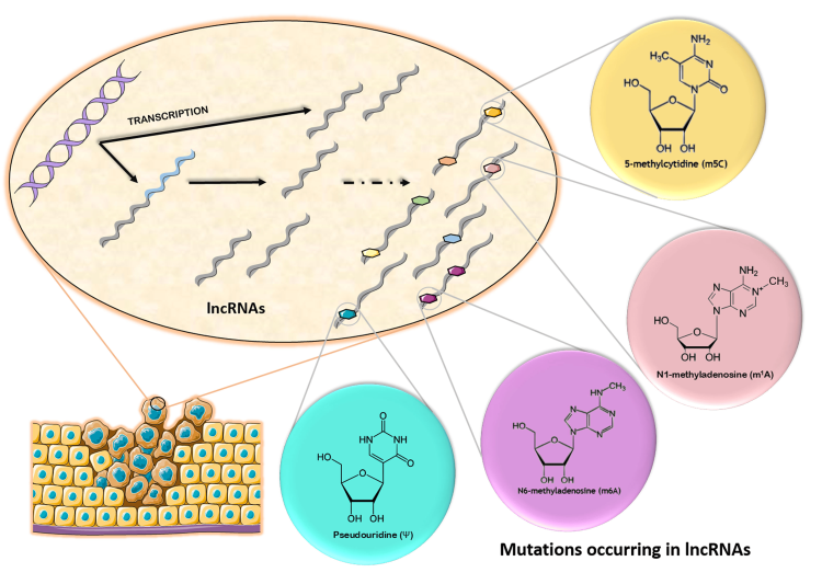
At the heart of this research lies the ribosome, the cellular machineries responsible for protein synthesis. Traditionally, ribosomes were believed to share a uniform structural design throughout the body. However, the team uncovered significant variations in chemical modifications across different tissue types, developmental stages, and disease states. These modifications affect ribosomal RNA (rRNA), ultimately altering ribosomal functionality.
In their investigation, the team analyzed rRNA from various organs of both humans and mice, including the brain, heart, liver, and testis. They discovered that each tissue displays a distinct rRNA modification pattern, termed the “epitranscriptomic fingerprint.”
Unique Signatures of Cancer Cells
These unique signatures on the ribosomes can indicate the source of the cells, akin to how each tissue leaves its own address label on cellular structures. In studies involving tumor samples from cancer patients, the researchers identified specific “fingerprints” associated with lung cancer and testicular cancer.
Focusing more intently on lung cancer, the team collected samples of normal and affected tissues from 20 patients diagnosed with stage I or II lung cancer. They confirmed that cancer cells exhibited a deficiency in rRNA modifications. Using this information, they trained an algorithm capable of accurately differentiating between samples based on their unique molecular fingerprints. This approach showed remarkable accuracy in distinguishing lung cancer from healthy tissue, addressing a profound need since most lung cancer cases are diagnosed at advanced stages. The new method holds potential for earlier detection, granting patients more time for effective treatment.

Advancements in Sequencing Technology
The breakthrough is rooted in advancements in nanopore direct RNA sequencing technology, which allows for real-time analysis of rRNA molecules and their modifications. The significant advantage of nanopore sequencing lies in its use of small, portable sequencing devices that can easily fit in one hand. By placing biological samples into the device, it captures and analyzes RNA molecules instantaneously.
The research demonstrates that scanning only about 250 RNA molecules from tissue samples can effectively differentiate cancerous cells from normal cells, showcasing the immense potential of nanopore sequencing technology.
A New Perspective on Cancer Diagnostics
This study not only challenges conventional beliefs about ribosomal consistency but also introduces a novel approach to cancer detection. As each type of cancer unveils its unique “epitranscriptomic fingerprint,” the foundation for developing quick, non-invasive diagnostic tools emerges. In the future, clinicians may even achieve the aspiration of detecting cancer through mere blood samples, significantly minimizing physical invasiveness for patients while enhancing the likelihood of early detection.


Discussion about this post